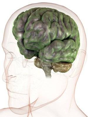
Computed tomography of the head as a method of in vivo imaging of the brain and its surrounding structures provide valuable information in the diagnosis and management of patients neurological and surgical patients. Knowledge of the location and nature of the pathology allows to choose correct tactics of treatment and to rationally plan the surgery.
When is computed tomography of the head
The study indicated for diagnosis when referring the patient to a relevant specialist. Imaging of the head has a high information content and resolving power method can be used for differential diagnosis of diseases with similar symptoms.
In any cases assigned to the study:
- traumatic head injuries, intracranial hemorrhage;
- diagnosis of ischemic and hemorrhagic strokes in the first hours after their occurrence;
- suspicion for the development of space-occupying lesions of the brain – tumors, cysts, abscesses;
- diseases of eyeball and orbit;
- inflammatory processes, neoplasms of the paranasal sinuses;
- acute and chronic inflammatory diseases of the head, encephalitis, meningitis, internal otitis, neuritis of cranial nerves;
- diagnostics of the dentition and facial skeleton;
- headache with unknown cause, lasting several months;
- sudden disturbances of vision, hearing, consciousness;
- suspected atrophic processes in the brain.
Head CT is performed in cases when the patient has contraindications to similar methods of study. In the process of treatment of oncological diseases, after operations method is used as a control to assess the effectiveness of medical intervention.
CT scan of the head fails with the following groups of patients:
- pregnant at any time;
- children under 18 years of age;
- mental illness with excessive excitation of the patient – the procedure can be under General anesthesia;
- the contrast material is contraindicated for pregnant women, lactating women, patients with renal and hepatic insufficiency, severe forms of hyperthyroidism in the presence of Allergy to iodine.
In each clinical case, the possibility of CT of the head is determined by the radiologist. If the benefit to the patient outweighs the potential harm, the study is conducted in the presence of contraindications.
Results
The results of the scan is represented by a series of layered images reflecting the structure of the soft tissues of the head and bones of the skull. After analyzing the images, the radiologist makes a report, which reflects all the features and pathological changes of the study area.
Normal in the pictures there is no area of pathological density, evidence of tumors, hemorrhage, or inflammatory process. For example, areas of hemorrhage appear as bright spots, tumors characterized by active accumulation of contrast medium and swelling of the adjacent brain. On the basis of the received images to construct a three-dimensional model and to estimate the spatial location of the pathological focus.
All the pictures the patient receives the electronic medium. Finally, the radiologist describes the revealed pathology, but is not a diagnosis. Depending on the nature of the violations, after a head CT should consult a neurologist, ENT, ophthalmologist, surgeon or oncologist. The specialist will deliver an accurate diagnosis and determine the tactics of treatment.
However, to get the results of the study need to choose a clinic for its passage. On the website https://mrt-v-spb.ru/mrt-golovy/ posted medical centers, providing services for the diagnosis of diseases of the head. Compare medical centers for the cost survey, rating, availability of shares, working hours, reviews and reputation of the clinic. If necessary, the patient can call the number 8 (812) 385-77-56 for appointment.